SCIENCE EPISODE
They Found an Aneurysm In My Brain: My MRI Story
I didn’t have a panic attack, but I just left the room — I just left my body. I was like, “What is going on? I thought you were going to tell me that I was perfectly fine and healthy, and you’re telling me…”
Hello angels, and welcome back to the Glucose Goddess Show. My name is Jessie Inchauspé, your favorite French biochemist — I think I can say that.
Today I wanted to do a sort of special episode: I wanted to talk about something personal that happened to me, in case it’s of interest and helps you in your health journey.
About a year and a half ago, I decided to have a full-body MRI scan. The reason was to make sure there was no early cancer in my body; I thought it could give me peace of mind, because so many people around me are getting these diseases younger and younger. I went to one of those companies that do full-body MRI scans and, let me tell you, the results were not what I expected — kind of crazy.
First, how do MRI machines work? They’re massive machines that look inside your body using magnets and radio waves. I was lying in the MRI machine, watching a documentary, while the machine worked around my body. An MRI is a big magnet — compared to a fridge magnet, it’s about 5,000 times stronger. Importantly, you can’t turn a fridge magnet on and off, but you can turn an MRI magnet on and off. These magnets are so powerful that if you were in the machine with your phone in your pocket, the phone would fly to the magnet because anything metallic is attracted almost instantly. Your body is made of trillions of atoms, many of them hydrogen. Once you get inside the big MRI magnet, it makes those hydrogen atoms align in the same direction. This is not dangerous; you don’t feel it, it has no consequences. Once the atoms are lined up, the MRI sends a radio wave through your body that gently nudges them out of alignment; as they realign, the MRI listens for the echoes. Different tissues — organs, bone, muscle — give different echoes, and the machine uses them to recreate images inside your body. It’s very cool technology and, unlike X-rays, it’s not harmful, which is why I was excited to try it. I was in the machine, having a great time, thinking, “For sure I’m super healthy.”
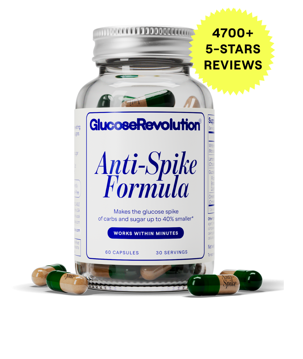
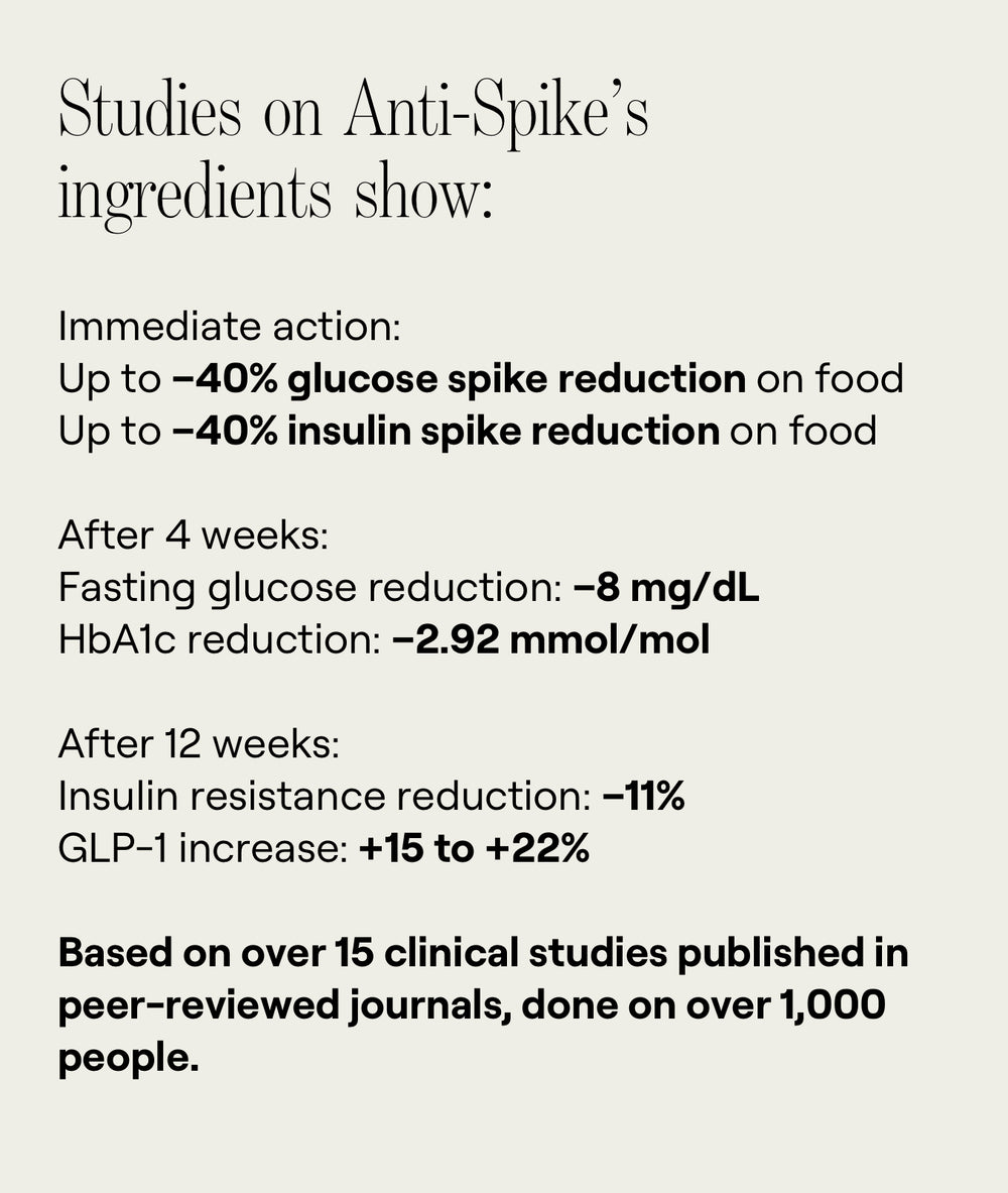
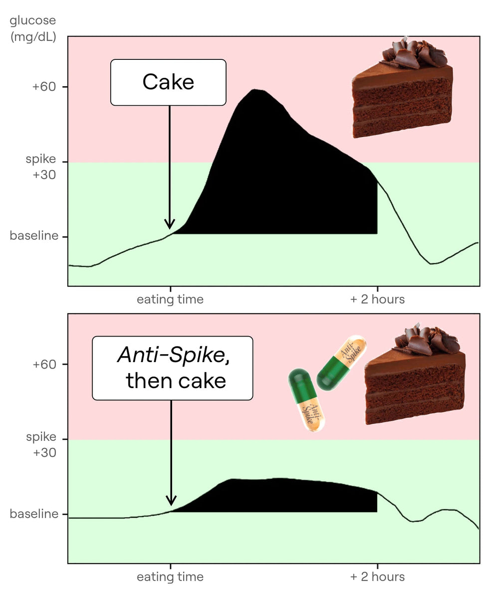
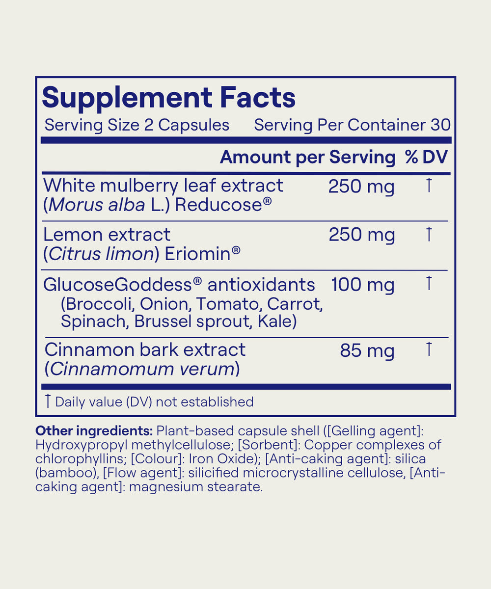
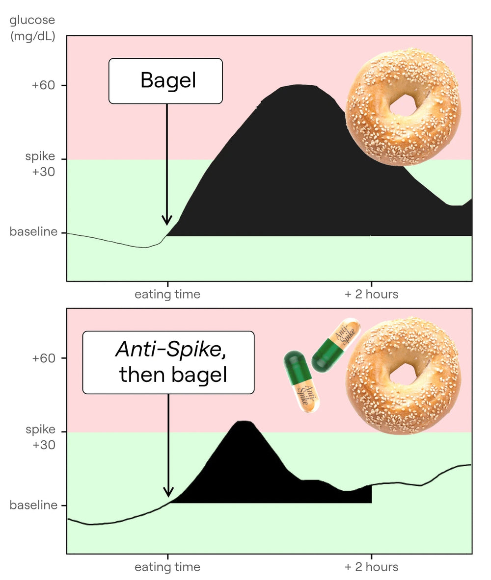
A few weeks later, I got a phone call from the doctors. (You guys know I’ve been doing my glucose hacks religiously — they’re the foundation of my dietary habits — and adding the molecules in Anti-Spike has allowed me to get to the next level: one, bloating — I feel so much better; two, energy levels — super consistent, eagle energy all day; three, sugar cravings — I love sugar, but Anti-Spike has given me a superpower over cravings. I know these natural molecules will help my long-term fasting glucose and fasting insulin, key to physical and mental health and healthy aging. Go to anti-spike.com to see the science, testimonials from thousands who’ve tried it, and to order your own Anti-Spike Formula bottle.)
First image they showed me: my brain in the MRI. Around my teeth area it’s dark, like a cloud — I have a permanent retainer that messes with the MRI signal; any metal causes this kind of shadow. Then things got scary. The doctor said, “We found something really interesting.” I thought, “Cool, interesting sounds good.” He said, “We found an aneurysm in your brain.” Cue all my blood leaving my body. I was in shock: “An aneurysm? I’ve only heard that they kill people.” He said they found a 4 mm aneurysm in my left internal carotid artery, basically behind my eyes. I was terrified. I didn’t have a panic attack, but I mentally checked out: “I thought you’d tell me I’m perfectly fine and you’re telling me there’s an aneurysm in my brain. Does that mean I’m going to die? Have a stroke? Is this dangerous? Can we do anything?” Here’s an image of it — a small, 4 mm bulge. As a healthy 30-year-old, I never thought they would find anything. Cold shower. First lesson: if you do a scan, be prepared — hopefully nothing shows, but something can.
They also showed me a video — I’ll point an arrow — of the blood vessels in the middle of my brain with the little aneurysm on one of them. Honestly, I was almost passing out from stress. The doctor was like, “We found this aneurysm; okay, let’s move on to your metabolism.” He also said, “This is good — finding an aneurysm is like Christmas for a doctor.” I thought, “What do you mean Christmas? I’m having an anxiety attack.” He explained they love to find these things because they’re usually benign and monitoring can prevent issues. Personally, it was rough; I was stressed for months as I worked through follow-up and what to do. I thought I had something in my brain that could rupture at any moment. An aneurysm is essentially a little malformation — a bulge in a blood vessel — which makes the vessel weaker; if it ruptures, you bleed into the brain, which can be very bad: death, lifelong consequences. A normal vessel is smooth; with an aneurysm you see that bulge. As I calmed down and read the research, I realized unruptured aneurysms — what I have — are really common: about 1 in 30 people worldwide (~3%) have an unruptured aneurysm, and most never find out because they don’t get full-body MRIs. If it never ruptures, you’ll never have issues; if it ruptures, that’s where problems start. While ~3% have an unruptured aneurysm, only ~0.001% of people will ever have a ruptured aneurysm — reassuring, though I still wondered: “Is mine bad? Big? Risky? How long has it been there?” About 85% of all unruptured aneurysms are in the Circle of Willis — the set of blood vessels in the center of the brain — exactly where mine is. With that info and after processing the freak-out, I sought follow-up care. I found a specialist and showed him my MRI. First reaction: “Why did you do a full-body MRI?” Then he said the next step was an angiogram to get a more precise image, because MRI isn’t that precise. They inserted a small catheter in my wrist, threaded it up to my brain, released a dye briefly, and took an X-ray at the same time. The photo they got is very detailed — much more than the MRI.
The good news: my aneurysm was small and benign enough that it’s better to leave it than to embolize it (placing a little coil to block it off). The risk of surgery was greater than the risk of rupture. About four months after the MRI, I had this result. I felt better, still nervous; I felt truly better a year later after a follow-up MRI showed it hadn’t grown. Overall, a stressful experience. Honestly, I’m not sure I’m happy I know — maybe I’d have preferred not to — because the stress was hard to move through.
But I did learn one thing we should all remember: the first symptom of a ruptured aneurysm is a very, very bad, intense headache. If I suddenly have the worst headache of my life, I need to rush to the ER.
That’s my takeaway. I wanted to share in case it’s helpful. I’m curious: would you want to know, or not? Tell me in the comments — I want to know how you feel. I’m still not really sure. Okay, I’ll see you next time. Lots of love.
 Anti-Spike Supplement
 The Recipe Club
 Course
 50 Breakfast Recipes
 50 Veggie Starter Recipes
 20 Vegan Recipes
 20 Gluten-Free Recipes
 The 10 Glucose Hacks
 Vinegar
 Alcohol
 Fasting
 GLP-1
GLP-1
 What to Eat Before & After Exercise
 Protein
 PCOS
PCOS
 Menopause
 My MRI Story
 Breakfast
 Supplements
 Clothes on Carbs
 Eggs & Cholesterol
 Chocolate
 Food Labels
 Veggie Starters
 Move After Eating
 Why Glucose Matters
 Glucose Revolution
 The Glucose Goddess Method
 9 Months That Count Forever


